Thursday, January 31, 2013

Pyogenic Granuloma


Description:
- Pyogenic Granuloma is a red, nodular overgrowth of granulation tissue that arises from the mucosal or skin surface.
- Known as a "Eruptive hemangioma", "Granulation tissue-type hemangioma", "Granuloma gravidarum", "Lobular capillary hemangioma", "Pregnancy tumor", and "Tumor of pregnancy".
- Most commonly, the lesion appears in the gingiva, and can also appear in lips, tongue, buccal
mucosa, palate, vestibule and edentulous areas, where the interdental papilla of the anterior maxilla is most common site for the lesion.
- The Pyogenic Granuloma bleeds easily and some cause mild pain.
- The Pyogenic Granuloma commonly develop during pregnancy, and is called "Granuloma Gravidarum"
or "Pregnancy Tumor".

Etiology:
- mild trauma, infection and hormonal disturbances are prominently mentioned, but the stimulus that provokes this overgrowth of granulation tissue is unknown.

Prognosis:
- Good

Treatment:
- Conservative excision.
- They may recur.
- Recurrent bleeding in either the oral or nasal lesions may necessitate excision and cauterization.
- If the lesion appears during pregnancy, it may heal spontaneously.

Differential Diagnosis:
- Peripheral Giant Cell Granuloma.
- Peripheral Ossifying Fibroma.

-------------------------

This Article has been Edited By :: World Of Dentistry :: TEAM
For any questions and Suggestions please don't be hesitate to feedback us.

Yours,
:: World Of Dentistry :: TEAM

Thursday, January 24, 2013

Cysts of the Jaw

- Description:
Cysts are usually painless when small and can cause significant bone loss before presenting clinically.
Cysts arising from odontogenic origin (odontogenic epithelium) account for most cysts of the jaws.

- Definition:
A cyst is a pathological cavity with fluid, semi-fluid or gaseous contents, which is not created by accumulation of pus. It is frequently lined by epithelium.

- Typical features of jaw cysts:
Most jaw cysts behave similarly—they grow slowly and expansively.
They differ mainly in relationship to teeth and radiographic features.
They form sharply-defined radiolucencies with smooth borders and are frequently a chance radiographic finding.

- Clinical Features:
- Noticeable swelling initially smooth bony hard lump with normal overlying mucosa, but as bone thins through resorption; cyst may show through as bluish fluctuant swelling (compressible).
- Discharge into mouth
- Pain due to secondary infection
- Fluid may be aspirated and thin-walled cysts may be transilluminated

- Classification of cysts of the jaws:

Odontogenic cysts:

1- Developmental:
- Gingival cyst of infants (arise from remnants of dental lamina)
- Dentigerous (follicular) cyst (arise in bone around crown of unerupted tooth)
- Eruption cyst (often burst spontaneously, rarely require excision)
- Odontogenic keratocyst (see radiograph on left, arise from dental lamina, multilocular, 60% recur, Gorlin-Goltz syndrome)
- Gingival cyst of adults (arise from epithelial rests in gingivae)

2- Inflammatory (55% of all cysts – epithelial rests of Malassez)
- Radicular cyst (= apex of non-vital tooth, see radiograph on right)
- Lateral Periodontal cyst (= lateral aspect of vital tooth)
- Residual cyst (cyst remains following removal of non-vital tooth)

3- Neoplastic
- Ameloblastoma (locally malignant, often multilocular, excision with wide margin of normal tissue, see radiograph below)
- Calcifying epithelial odontogenic (Pindborg, mixed radiolucency, locally invasive)

Non-Odontogenic Cysts:

- Nasopalatine Duct Cyst (Incisive Canal Cyst) - (Care not to confuse with anterior palatine fossa on x-ray)
- Nasolabial cyst (Nasolabial fold swelling – distorts nostril)
- Median Palatine, Median Mandibular Cysts

Cysts without Epithelial Lining:

- Solitary Bone Cyst (Simple Bone Cyst) - (arches up between roots of teeth, Rx.=allow bleeding into cavity)
- Aneurysmal Bone Cyst (multilocular, 10-20yrs, very expansile, blood-filled spaces, giant cells)

- Diagnostic tests:
- Radiographs - intra and extra-oral views
- Vitality testing of associated teeth.
- Aspiration and analysis of fluid contents.
- Biopsy of lining, provides definitive diagnosis

- Diagnosis through Cyst contents:
- Straw-coloured fluid containing cholesterol crystal indicates most Radicular cysts
- Air, some blood or serosanguious fluid indicates Solitary Bone cysts
- Blood indicates Aneurysmal Bone cyst
- Pus indicates infected cyst
- Pale, yellow fluid containing keratin indicates Odontogenic Keratocyst

- Management:
1. Enucleation with primary closure is most common, and generally the treatment of choice. It consists of removing the cyst lining from the bony walls of the cavity and repositioning the access flap. Any relevant dental pathology is treated at the same time.
2. Enucleation with packing and delayed closure is used when badly infected cysts, particularly very large ones, are unsuitable for primary closure. Pack with Whitehead’s varnish.
3. Enucleation with primary bone grafting
4. Marsupialization—opening of the cyst to allow continuity with the oral mucosa. Healing is slower than with enucleation and a cavity persists for some time.

---------------------------

 This Article has been Edited By :: World Of Dentistry :: TEAM
For any questions and Suggestions please don't be hesitate to feedback us.

Yours,
:: World Of Dentistry :: TEAM

Sunday, January 20, 2013

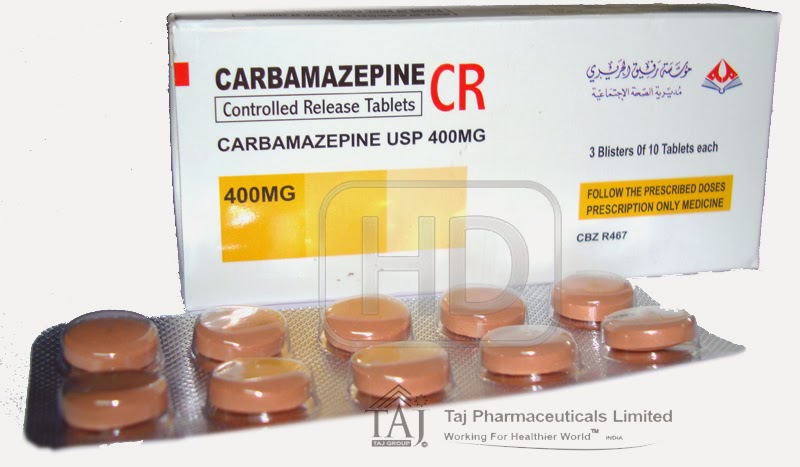
Carbamazepine


- Description:
Carbamazepine is in a group of drugs called anticonvulsants.
It works by decreasing nerve impulses that cause seizures and pain.
Carbamazepine is used to treat seizures and nerve pain.
It may interfere with contraceptives
Do a FBC soon after starting carpamazepine
Blood Dyscrasias usually occur in the first 3 months.
Dose: 100-200 mg once a day, gradually increased to 200 mg twice a day, with maximum of 1600 mg of separated doses per day.

- Indications:
1- Trigeminal Neuralgia
2- Glossopharyngeal Neuralgia
3- Diabetic Neuropathy
4- Bipolar Disorder

- ContraIndications:
1- Drug Allergy
2- Patients with atrioventricular conduction defects.
3- porphyria

- Extreme Percution:
1- Pregnant Females.
2- Patients on MAOIs (Monoamine Oxidase Inhibitor)
3- Liver Failure.

- Side Effects:

1- Major:
- a red, blistering, peeling skin rash;
- fever, sore throat, body aches, flu symptoms;
- easy bruising or bleeding, unusual weakness;
- white patches or sores inside your mouth or on your lips;
- feeling short of breath, even with mild exertion;
- swelling of your ankles or feet;
- nausea, stomach pain, low fever, loss of appetite, dark urine, clay-colored stools, jaundice (yellowing of the skin or eyes);
- urinating less than usual.

2- Minor:
- feeling unsteady;
- mild nausea, vomiting, diarrhea, constipation, stomach pain;
- confusion, headache, blurred vision;
- feeling agitated or depressed;
- ringing in your ears;
- dry mouth, swollen tongue; or
- joint or muscle pain, leg cramps.

------------------------------

This Article has been Authored By :: World Of Dentistry :: TEAM
For any questions and sugesstions please don't be hesitate to feedback us.

Yours,
:: World Of Dentistry :: TEAM
Condensing Osteitis


- Description:
It's a periapical inflammatory diseases, that results from reaction to infection, which is periodontal and not dental.
There would be bone production rather than bone destruction.
The most common site is near premolars and molars.
The Lesion appear as an radio-opacity in the periapical area hence the sclerotic reaction.
The sclerotic reaction result from good patient resistance and a low degree of virulence of the offending bacteria.
The associated tooth is carious or contains a large restoration.

- Synonyms:
1- Garre's Disease (described by Dr. Carl Garré in 1893).
2- Sclerosing Osteitis.

- Etiology:
Infection of periapical tissues of a high immunity host by organisms of low virulence.

- Treatment:
The offending tooth should be tested for vitality of the pulp, if inflamed or necrotic, then endodontic treatment is required, while hopeless teeth should be extracted.

- Prognosis:
if the offending tooth is extracted, the area of condensing osteitis may remain in the jaws indefinitely, and is termed osteosclerosis or bone scar.

- Differential Diagnosis:
1- Idiopathic osteosclerosis.
2- cementoblastoma.
NOTE: An abnormal result with pulp testing strongly suggests condensing osteitis and tends to rule out osteosclerosis and cementoblastoma.

------------------------------

This Article has been Authored By :: World Of Dentistry :: TEAM
For any questions and sugesstions please don't be hesitate to feedback us.

Yours,
:: World Of Dentistry :: TEAM

Thursday, January 10, 2013

Medications in Pregnancy

    When dealing with the pregnant patients, a special protocols should be followed, regarding the seat position, the visit duration, the dental treatment accepted, the anesthesia, the visit time, the period, ... etc.
    Here we are pointing to the "Drugs" are to be prescribed to the pregnant female which can be categorized as the following pattern, but N.B those recommendations.
    - General Recommendations:
    A- Avoid medications if possible in first trimester
    B- Limit use to safe, short-acting, non-combination drugs
    C- Topical medications are preferred over systemic agents
    D- Use the lowest effective dose of a medication
    ---------------------------
    - Categories of Drugs Prescribed to Pregnant Females:
    1- Class A
    - No risk in controlled human studies
    - Examples: Acetaminophen (Tylenol) - Pyridoxine (Vitamin B6)
    2- Class B
    - No risk in controlled animal studies
    - Examples: Amoxicillin - Cephalosporin antibiotics
    3- Class C
    - Small risk in controlled animal studies
    - Examples: Codeine - Dicloxacillin
    4- Class D
    - Strong evidence of risk to the human fetus
    - Examples: Coumadin - Valium
    5- Class X (Never to be used in Pregnancy)
    - Very high risk to the human fetus
    - Examples: Xanax - Accutane
    ---------------------------
    Analgesics Related to Pregnant Female:
    Antibiotics Related to Pregnant Female:
    ---------------------------
    - References:
    - Briggs (1998) Drugs in Pregnancy and Lactation, 5th ed
    - Larimore (2000) Prim Care 27(1):35-53
--------------------------------

This Article has been Authored By :: World Of Dentistry :: TEAM
For any questions and sugesstions please don't be hesitate to feedback us.

Yours,
:: World Of Dentistry :: TEAM

Tuesday, January 1, 2013

Antibiotics in Pregnancy


Class B: No risk in controlled animal studies

A- Antifungal Topical Agents
Nystatin (Mycostatin)

B- Antiparasitic agents
Metronidazole or Flagyl (after first Trimester)
Avoid single dose therapy
Praziquantel - not in Briggs
Permethrin (topical) - not in Briggs (1998)

C- Anti-Tuberculosis agents
Ethambutol
Didanosine - not in Briggs (1998)

D- Antiviral agents
Nelfinavir - not in Briggs (1998)
Ritonavir
Saquinavir
Famciclovir
Valacyclovir

E- Antibiotics
All Cephalosporin Antibiotics (except Moxalactam)
All Erythromycin except Erythromycin Estolate
Azithromycin (Zithromax)
All Penicillin Antibiotics
Clindamycin
Macrodantin (before third trimester)
Sulfa antibiotics (before third trimester)

-------------------------------

Class C: Small risk in controlled animal studies

A- Antifungal agents
CDC recommends only Topical Antifungal in pregnancy
Avoid Antifungals in first trimester if possible
Terbinafine (Lamisil) - not in Briggs (1998)
Clotrimazole (Mycelex, Lotrimin)
Butoconazole (Femstat)
Miconazole (Monistat)
Amphotericin B
Fluconazole (Diflucan)
No fetal adverse effects seen in one study
King (1998) Clin Infect Dis 27:1151-60
Itraconazole (Sporanox)
Ketoconazole (Nizoral)
Teratogenic and Embryotoxic in animals
Griseofulvin
Teratogenic and Embryotoxic in animals

B- Antimalarial agents
Mefloquine (Lariam)
Chloroquine
Primaquine

C- Antiparasitic agents
Albendazole - not in Briggs (1998)
Ivermectin - not in Briggs (1998)
Mebendazole
Pentamidine
Thiabendazole
Pyrantel

D- Anti-Tuberculosis agents
Dapsone
Isoniazid (INH)
Pyrazinamide
Rifampin

E- Antiviral agents
Lamivudine
Stavudine
Zalcitabine
Zidovudine
Delavirdine - not in Briggs (1998)
Nevirapine
Indinavir
Cidofovir
Foscarnet
Ganciclovir
Acyclovir
Amantadine
Rimantadine
Interferon alpha

F- Antibiotics
Imipenem-Cilastin
All Fluoroquinolone antibiotics
Clarithromycin (Biaxin)
Pediazole - not in Briggs (1998)
Sulfisoxazole - not in Briggs (1998)
Trimethoprim
Vancomycin
Chloramphenicol
Gentamicin

-------------------------------

Class D: Strong evidence of risk to the human fetus

A- Antiparasitic agents
Metronidazole or Flagyl (First Trimester)
1- New evidence suggests first trimester safety
2- Burtin (1995) Am J Obstet Gynecol 172:525-9

B- Antibiotics
- Amikacin (Class D per manufacturer)
- Kanamycin
- Streptomycin
- Tobramycin (Class D per manufacturer)
- Sulfa (Third Trimester)
- All Tetracycline antibiotics (Doxycycline, Tetracycline and Minocycline)
- Erythromycin Estolate (llosone) - Due to hepatotoxicity in pregnant women
- Macrobid and Nitrofurantoin (Third Trimester)
1- Do not use either of these past 38 weeks
2- Can cause Hemolytic Anemia in newborns
3- Related to immature liver and G6PD Deficiency

C- Vaccines
Yellow Fever Vaccine
-------------------------------

Class X: Very high risk to the human fetus

A- Antimalarial agents
- Quinine

B- Antiviral agents
- Ribavirin
- Rebetron - not in Briggs (1998) 

C- Vaccines
- MeaslesVaccine
- Mumps Vaccine
- RubellaVaccine
- Small PoxVaccine
- TC-83 Venezuelan Equine EncephalitisVaccine
- Varicella Vaccine
1- Risk if vaccinated within 4 weeks of conception
2- Theoretic risk only; not an indication for EAB

--------------------------------

This Article has been Authored By :: World Of Dentistry :: TEAM
For any questions and sugesstions please don't be hesitate to feedback us.

Yours,
:: World Of Dentistry :: TEAM