Saturday, December 28, 2013

Enucleation of Oral Cysts


- Description:
- Enucleation is the process by which the total removal of a cystic lesion is achieved.

- By definition, it means a shelling-out of the entire cystic lesion without rupture.

- A cyst lends itself to the technique of enucleation because of the layer of fibrous connective tissue between the epithelial component (which lines the interior aspect of the cyst) and the bony wall of the cystic cavity.

- This layer allows a cleavage plane for stripping the cyst from the bony cavity and makes enucleation similar to stripping periosteum from bone.

- Enucleation of cysts should be performed with care, in an attempt to remove the cyst in one piece without fragmentation, which reduces the chances of recurrence by increasing the likelihood of total removal.

- In practice, maintenance of the cystic architecture is not always possible, and rupture of the cystic contents may occur during manipulation.

- The Periapical (i.e., Radicular) cyst is the most common of all cystic lesions of the jaws.

- After removing the Cyst, the bony cavity fills with a blood clot, which then organizes over time.

- Radiographic evidence of bone fill will take 6 to 12 months after removing the cyst.

- Jaws that have been expanded by cysts slowly remodel to a more normal contour.

- If the primary closure should break down and the wound dehiscence occurs,
1- Recall visits for irrigation every 3-4 days.
2- Strip gauze lightly impregnated with an antibiotic ointment should be gently packed into the cavity.
3- in 3-4 days, then cavity will start to be filled with granulation tissue.

- Indications:
1- Enucleation is the treatment of choice for removal of cysts of the jaws and should be used with any cyst of the jaw that can be safely removed without unduly sacrificing adjacent structures.
2- Small Cysts
3- Away from Important Anatomical Structures.
4- Not at the bone margins (may lead to jaw fracture)
5- Not associated with teeth, roots or important structures.

- Advantages:
1- The main advantage to enucleation is that pathologic examination of the entire cyst can be undertaken.
2- Another advantage is that the initial excisional biopsy (i.e., enucleation) has also appropriately treated the lesion.
3- The patient does not have to care for a marsupial cavity with constant irrigations.
4- Once the mucoperiosteal access flap has healed, the patient is no longer bothered by the cystic cavity.

- Disadvantages:
If any of the conditions outlined under the section on indications for marsupialization exist, enucleation may be disadvantageous. For example,
1- Normal tissue may be jeopardized,
2- Fracture of the jaw could occur,
3- Ddevitalization of teeth could result, or associated impacted teeth that the clinician may wish to save could be removed.

- Technique: (Small Cysts)
1- Remove all possible irritants before starting with enucleation procedure (ex: subgingival and supragingival scaling and stains).
2- Administration of Local Anesthetic Solution, Lignocaine 2% with adrenaline 1:200,000 is used.
3- Nerve Block associated with an infiltration is preffered.
4- Flap design is picked accoring to the possition of the lesion (ex: crevicular incision with a 2 vertical releasing incisions to give a good access and visibility to the lesion).
5- Use Mucoperiosteal Elevator to raise the flap. (care not to tear tissues)
6- Use burs to remove all thin, resorbed, infected and soften bone. (care not to remove excessive bone or damage to adjacent roots and anatomical structures)
7- Remove the entire cyst carefully. (care not to tear or punch the cyst lining).
8- A Piece of rolled gauze is hold by a hemostat and inserted between the cavity and the lining.
9- The Cavity is then rinsed with Saline and Betadine.
10- The bony edges of the defect should be smoothed with a bone file before closure.
11- Reposition the flap, and suture it then with the proper suture type and suturing technique. (Vertical Mattress interrupted is preffered using a Black Braided Silk Suture type).

- The use of antibiotics is unnecessary unless the cyst is large or the patient's health condition warrants it.

- If, on the other hand, the tooth is restorable, endodontic treatment followed by periodic radiographic follow-up will allow assessment of the amount of bone fill.

- When extracting teeth with periapical radiolucencies, enucleation via the tooth socket can be readily accomplished using curettes when the cyst is small. (Caution is used in teeth whose apices are close to important anatomic structures, such as the inferior alveolar neurovascular bundle or the maxillary sinus, because the bone apical to the lesion may be very thin or nonexistent).

- Technique: (Large Cysts)
1- A mucoperiosteal flap may be reflected and access to the cyst obtained through the labial plate of bone, which leaves the alveolar crest intact to ensure adequate bone height after healing.
2- Open an osseous window, and begin to enucleate the cyst.
3- A thin-bladed curette is a very suitable instrument for cleaving the connective tissue layer of the cystic wall from the bony cavity.
4- The largest curette that accommodates the size of the cyst should be used.
5- The concave surface should always be kept facing the bony cavity—the edge of the convex surface performs the stripping of the cyst.
6- try to keep in cyst margins intact to facilitate its removal.
7- The cyst separates more readily from the bony cavity when the intracystic pressure is maintained.
8- Nerves and Vessels might be embedded or pushed to one side of the large cyst, thus, should be carefully handled.
9- Inspected the bony cavity for remnants of tissue.
10- Irrigating and drying the cavity with gauze will aid in visualizing the entire bony cavity.
11- Residual tissue is removed with curettes.
12- The bony edges of the defect should be smoothed with a bone file before closure.

-------------------------------

This Article has been Edited By :: World Of Dentistry :: TEAM
For any questions and suggestions please don't be hesitate to feedback us.

Yours,
:: World Of Dentistry :: TEAM

Thursday, December 5, 2013

Medical Complications in Pregnancy


* Many changes occurs to female body during the pregnancy period, due to the hormonal changing:

* Some of the medical complications of pregnancy can include:

1- Hypertension:
- Very common and blood pressured becomes 140/90 mm Hg or greater.
- Hypertention is a dangerous complication that may effect both fetus and mother.
- Hypertensive pregnant women must rest as much as possible and treated with anti-hypertensive treatment.

2- Blood Hypercoagulability:
- Excessive thickening of blood, which can lead to venous thrombosis (occlusion of blood vessels),
- Disseminated Intravascular Coagulopathy (A rare, life-threatening condition that prevents a person’s blood from clotting normally).

3- Anemia:
- A deficiency of red blood cells. 
- Expansion of the blood volume may cause an apparent anemia but in about 20%.
- True anemia develops, mainly because of fetal demands for iron and folate.
- Pregnancy may complicate a pre-existing Anemias, especially the Sickle Cell Anemia.

4- Supine Hypotension Syndrome.
- In later pregnancy up to 10% of female patients may become hypotensive, if laid in supine position for 3 to 7 mins, when the gravid uterus compresses the Inferior Vena Cava and impedes blood return to the heart and may lead to syncope.

---------------------------------

This Article has been Edited By :: World Of Dentistry :: TEAM
For any questions and suggestions please don't be hesitate to feedback us.

Yours,
:: World Of Dentistry :: TEAM

Saturday, November 23, 2013

Phlebitis and Thrombophlebitis


Definition:
Phlebitis means inflammation of a vein. TP ( Thrombophlebitis ) is the term used when a blood clot in the vein causes the inflammation. TP usually occurs in leg veins, but it may occur in an arm. The thrombus (clot) in the vein causes pain and irritation and may block blood flow in the veins. Phlebitis can occur in both the surface (superficial) or deep veins.

Sign and Symptoms:
1- Pain
2- Tenderness
3- Edema
4- Erythema
5- Streaking of The Limb

Ideal Treatment: 
1- Remove of the IV Catheter.
2- Elevate the Affected Limb.
3- Apply Warm, Moist Packs to the infected site.
4- Initiate IV Antibiotics.

-------------------------------

This Article has been Authored By :: World Of Dentistry :: TEAM
For any questions and suggestions please don't be hesitate to feedback us.

Yours,
:: World Of Dentistry :: TEAM

Monday, October 28, 2013

Oral Side-Effects of Chemotherapy


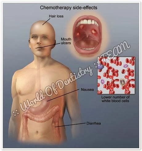
The most common oral complications observed after chemotherapy are mucositis, infections, neurological and dental alterations, dysgeusia, hyposialia and xerostomia (dry mouth), bleeding tendency, and the development of osteonecrosis. The soft tissues of the lips, the oral mucosa, tongue, soft palate and the pharyngeal mucosa are the most affected areas.

1- Mucositis:
- Mucositis is an inflammatory reaction of the mucosal membranes secondary to antineoplastic treatments such as radiotherapy (in 80% of the cases) and chemotherapy as treatment for solid tumors or lymphomas (in approximately 40-50%, particularly with the cytostatic agent 5-fluorouracil) or as conditioning treatment for bone marrow transplantation (in over 75% of the patients).
- Mucositis is regarded as a manifestation of leukopenia.
- The mechanism by which mucositis develops is not clear, though it is generally attributed to the fact that the oral mucosal cells have a relatively high mitotic rate, thereby establishing them as targets of the action of cytostatic agents.
- Mucositis usually appears 4-7 days after the start of highdose chemotherapy, and is of a self-limiting nature (provided Over-infection does not occur). It in turn disappears 2-4 weeks after the conclusion of cytotoxic chemotherapy.
- The drugs most often associated with the development of mucositis are doxorubicin, bleomycin, fluorouracil and methotrexate.
- Clinically, the condition manifests as erythema, edema or ulceration, with severe pain, bleeding and potential side effects such as xerostomia, the risk of both local (overinfection due to Candida) and systemic infection, malnutrition, fatigue, dental caries and gastrointestinal disorders over time.

2- Oral infections:
The main infectious processes are the following:

1. Bacterial infections: These are usually caused by gram-negative organisms. Signs of inflammation may be masked as a result of the underlying bone marrow suppression; consequently, oral hygiene protocols that reduce microbial colonization of the dentition and periodontium are important during the period of bone marrow suppression.

2. Fungal infections: Bone marrow suppression, oral mucosal lesions and salivary alterations contribute to the development of Candida albicans infection. The most common presentations are pseudomembranous candidiasis, followed by erythematous candidiasis. The treatment of these conditions involves the use of topical and/or systemic antifungal agents (Table 2) complemented with an antiseptic (chlorhexidine). The latter should be used at least 30 minutes before or after nystatin, since the combination of both may prove ineffective. In the case of more severe infections, the recommendation is a systemic antifungal such as fluconazole or ketoconazole.
The efficacy of such treatment is limited, and resistances may appear. In such cases intravenous amphotericin B or itraconazole via the oral route at a dose of 200-400 mg/day tend to be the drugs of choice.

3. Viral infections: In most cases, infections due to herpes simplex virus (HSV), varicella-zoster virus (VZV) and Epstein-Barr virus (EBV) are the result of the reactivation of a latent virus, while infections due to cytomegalovirus (CMV) can result from the reactivation of a latent virus or from a recently acquired virus.

- Infection due to HSV: The severity of the lesions increases drastically with the degree of immune suppression.
The treatment of HSV infection consists of the administration of 400-800 mg of aciclovir via the oral route 5 times a day or of 5-10 mg/kg via the intravenous route every 8-12 hours, for as long as the lesions persist.

- Infection due to VZV: Immune compromised patients may present involvement of several dermatomes, or alternatively the lesions may show a more generalized distribution, generally manifesting several weeks after the interruption of chemotherapy – in contrast to the situation with HSV.
A number of antiviral agents are used as treatment, depending on the degree of immune suppression of the patient and the resistances to these drugs (Table 2).

- Infection due to CMV and EBV: Lesions produced by CMV are characterized by the presence of multiple mild or moderate ulcerations with irregular margins. The initial lesions appear during the first periods of bone marrow regeneration and are characterized by nonspecific pseudomembranous ulcers covered by a fibrin exudate with a granulomatous base.
At present, ganciclovir is the treatment of choice for acute CMV infection. The risk of EBV infection usually manifests months after the interruption of myeloablative therapy used for transplant conditioning.

3- Neurotoxicity:
- A number of chemotherapeutic agents such as vincristine and vinblastine are able to cause direct neurotoxicity.
- Patients may experience deep and palpitating mandibular pain that tends to subside one week after concluding chemotherapy.
- A correct anamnesis is required, together with oral exploration and an X-ray study in order to distinguish such pain from pain of pulp origin.
- In some cases, dental hypersensitivity may appear weeks or months after the end of chemotherapy; in these cases, the topical application of fluoride or the use of a desensitizing toothpaste may help lessen the symptoms.

4- Dysgeusia:
- During chemotherapy, patients may experience an unpleasant metallic taste due to diffusion of the chemotherapeutic agent into the oral cavity.
- Dysgeusia as such initially manifests a few weeks after starting cytotoxic treatment, and is generally reversible within a few weeks.

5- Hyposialia and xerostomia:
- Hyposialia, attributable to the effect of chemotherapy upon the cells of the salivary glands, is transient and reversible.
- It appears particularly with the use of adriamycin, and can cause oral functional problems, especially in relation to speech and mastication. 
- These patients show alterations in the salivary components, with an increase in the levels of peroxidase and amylase, a reduction in total secreted immunoglobulins A and G, and the presence of the chemotherapeutic drug itself. all these factors favor the development of mucositis.
- As a result, patients should drink abundant water and use sugar-free sweets or chewing gum to increase salivation.
- In more moderate cases, sialogogues such as pilocarpine, bromhexine or bethanechol can be used.

6- Alterations in dental growth and development:
- Unlike radiotherapy, which only affects the cells within the irradiated zone, chemotherapy has a systemic effect.
- As a result, the developing odontogenic cells are susceptible to chemotherapy, even when far removed from the tumor site.

7- Bleeding tendency:
- Bleeding is due to alterations resulting from thrombocytopenia (in turn a consequence of bone marrow aplasia).
- Clinically, patients my present petechiae, ecchymosis, hematomas or diffuse bleeding.
- Rinses with 0.12% chlorhexidine avoid overinfection and can help eliminate the traces of blood, though caution is required in order not to alter the clots, since this could lead to further bleeding. 

8- Osteonecrosis:
- Osteonecrosis of the jaw (ONJ) is observed in patients treated with bisphosphonates (BPs).
- These drugs inhibit bone resorption and are administered via the intravenous route as treatment in application to bone metastases in cancer patients, in malignant hypercalcemia (tumorinduced hypercalcemia), or in patients with multiple myeloma – affording improved survival and quality of life.
- Although much less commonly, ONJ has also been observed in patients receiving treatment with oral bisphosphonates (used for the prevention and treatment of osteoporosis and in certain bone conditions such as Paget’s disease).
- The intravenous BPs most associated with ONJ are zoledronic acid (Zometa®) and pamidronate (Aredia®).
- Clinically, ONJ is typically characterized by pain (progressive and sustained, and sometimes requiring important analgesic doses to secure control – the patient being asymptomatic in the early stages).
- Since the treatment of ONJ is often unsatisfactory, management should aim to afford pain relief, control soft tissue and bone infection, and avoid or reduce the progression of bone necrosis.

-----------------------------------

This Article has been Edited by :: World Of Dentistry :: TEAM
For any suggestions please don't be hesitate to feedback us

:: World Of Dentistry :: TEAM

Monday, October 21, 2013

Root Canal Preparation


- Objectives of Root canal preparation:
1- Remove remaining pulp tissue
2- Eliminate microorganism
3- Remove debris
4- Shape the root canal
5- So that root canal system can be cleaned and filled

- Requirements of Root canal preparation:
1- Prepared canal should include the original canal
2- Apical constriction should be maintained
3- Canal should end in an apical narrowing
4- Canal should be tapered from crown to apex
5- Preparation should be undertaken with copious irrigation
6- The final length of the preparation should not be reduce by treatment

- Root canal preparation techniques:
According to Direction:
1- Apical to coronal
2- Coronal to apical

According to Type:
1.Standardized technique
2. Step back technique
3. Balanced forces technique
4- Step down technique
5- Double flared technique
6- Crown down pressure less technique

- Standardized technique
Can be use for straight tiny canal
Unsuitable for curve canals

Common problems:
1- Ledging
2- Zipping
3- Elbow formation
4- Perforation
5- Loss of working length

- Overcomes procedural errors of Standardized technique:
- Suitable for Slight to moderate curve canals
- Not suitable for severely curve canals
- Can be improve by: "Specific filing technique - Non-cutting tip - Flexible files"

- Steps of successful Root Canal Preparation:
1- Obtain a good pulp chamber cleaning.
2- Make sufficient deroofing for the pulp chamber area "ensure that files enter orifices in a straight manner"
3- Use GG to widen the orifices.
4- Insert the initial file which is "size 15 for centrals and premolars" and "size 10 for molars" Note: "size 8 may be useful for severely narrow canals ex: MB of the lower first molars"
5- Start to measure the full working length using the initial file and X-ray or by using the Apex Locator. Note: file size 10, 8 and 6 may not appear in the X-ray so that they may lead to misleading or interpretation for the working length, Note: It's better to have a good X-ray image with the length measured by Apex Locator to avoid false Apex Locator's readings ..
6- Start to widen canals to 3 files larger than the initial file to the full working length measured before, to create the good apical stop ... "The third file will be called the patency file".
7- Start to use the larger next file with a length 1 mm shorter than the previous one ..
8- Use 5 more larger files, every one 0.5 mm shorter the previous one.
9- Use the patency file between  files to avoid blockage of canals ...

10- Start to put the master cone and complete obturation ....

-----------------------------------

This Article has been Edited by :: World Of Dentistry :: TEAM
For any suggestions please don't be hesitate to feedback us

:: World Of Dentistry :: TEAM

Melkersson-Rosenthal Syndrome


- Definition:
Melkersson-Rosenthal syndrome is a rare neurological disorder characterised by recurring facial paralysis or palsy, swelling of the face and lips (usually the upper lip), and the development of folds and furrows in the tongue.

- Does it develop in childhood ... ?
Onset is in childhood or early adolescence.
After recurrent attacks (ranging from days to years in between), swelling may persist and increase, eventually becoming permanent. 
The lip may become hard and cracked with a reddish-brown discoloration. 
The tongue may develop furrows.

- Causes:
The cause of Melkersson-Rosenthal syndrome is unknown, but there may be a familial link although a gene has not yet been identified.

- Treatment:
Treatment is symptomatic and may include medication therapies with anti-inflammatory medication and corticosteroids to reduce swelling.
Occasionally surgery may be recommended for severe cases to relieve pressure on the facial nerves and to reduce swollen tissue, but its effectiveness has not been established.
Facial Rehabilitation is available from specialist centers where physiotherapists and speech and language therapists specialized in treatment for people with facial palsy. 
The treatment is similar to the treatment for Bell’s palsy although the causes of the two conditions are different.

- Prognosis:
Melkersson-Rosenthal syndrome may recur intermittently after it first appears.
The person will therefore experience intermittent facial weakness which can resolve as the nerve recovers.
It can become a long standing problem as with each attack recovery is less satisfactory.

-----------------------------------

This Article has been Edited by :: World Of Dentistry :: TEAM
For any suggestions please don't be hesitate to feedback us

:: World Of Dentistry :: TEAM

Saturday, September 14, 2013

Camphor Mono-Chlorophenol (CMCP)


Solution for Root Canal Disinfection

- Composition:p-Chlorophenol 35%, Camphor 65%

- Properties:PD Camphor Mono-Chlorophenol, also known as CMCP, is an active disinfectant for the treatment of infected root canals & periapical infections.

- Indication:- Disinfection after pulpectomy.
- Treatment of post-traumatic inflammation.
- Pulp dressing.

- How to use:- Dry the root-canal thoroughly.
- Complete the pulpectomy.
- Insert 1 or 2 drops of preparation into the root canal & allow few minutes for it to take effect.
- Remove any excess solution with a cotton pellet or an absorbent paper point.
- Remove gangrenous pulp tissue.
- Use CMCP as a dressing by inserting it into the root canal with a smooth broach.
- Obturate canals temporarily.
- Leave it ‘in situ’ for at least 3 days.
- Make sure the canal is completely disinfected after the period.
- If this is not the case, repeat the dressing before the final obturation.

- Storage:Store at room temperature between 5°C & 30°C, protected from direct light.

- Packaging:Bottle of 15 ml.

----------------------------------

This Article has been Edited by :: World Of Dentistry :: TEAM
For any suggestions please don't be hesitate to feedback us

:: World Of Dentistry :: TEAM

Friday, September 6, 2013

Gingival Hyperplasia


- Enlargement of gums or gingiva is called gingival hyperplasia in the dental terms.
- This occurs due to abnormal multiplication of gingival tissues due to various factors. 
- When an individual is diseased with gingival hyperplasia then the gums become swollen and enlarged. 
- If you are suffering from gingivitis there will be bleeding in the gums as well as tenderness. 
- The teeth get covered with the gums if the infection is too intense. 
- The patient is examined by a dentist and then the patient is sent for further examination if any further complications are seen.

---------------------------------

Causes:

There are several causes of gingival hyperplasia:

- Inflammatory and granulomatous disorders "like Hand-Schuller-Christian syndrome"
- Auto-immune disorders "like plasma cell gingivitis which is associated with allergic and collagenous nature"
- Neoplastic disorders "like acute leukemia and monocytic leukemia"
- Metabolic and storage disorders "like aspartylglycosaminuria, genetic disorders like Cross syndrome"
- Toxicity of drugs.
- Excess of deposition of drugs given to a patient. It is also called as Drug-Induced Gingival Overgrowth (DIGO).
- Immunosuppressant like Cyclosporin, anticonvulsants like valproate, phenytoin, phenobarbital, primidone and calcium channel blockers like nifedipine, verapamil and amlodipine.

Note: Cyclosporin and phenytoin toxicity are the important toxicities caused in a patient especially in case of dental plaque where these drugs get deposited on the tooth.

Causes of Gingival Enlargement is divided into five groups:
- Inflammatory Enlargement
- Enlargement due to systemic /genetic diseases
- Neoplastic enlargement
- False enlargement
- Drug induced enlargement
- Gingival Hyperplasia differential diagnosis

---------------------------------

Diagnosis:

- Mainly done by differential diagnosis.
- IFN-gamma-stimulated gene expression which is important to determine the presence of gingival hyperplasia in the body.

---------------------------------

Treatment:

The treatment of gingival hyperplasia is based on the type and cause of the gingival hyperplasia.
- Improving oral hygiene is the most important factor which will determine the cure rate of the disease.
- Maintain oral hygiene regularly to prevent any further spread of infection.
- Gingivectomy, a surgical procedure to remove the excess of gingival tissue is carried out to manage the condition.
- The drugs which cause toxicities and affect the gingival tissues can be replaced with alternative drug therapies. For instance, in case of immunosupression, cyclosporine can be replaced by tacrolimus which is has less affect in gingival hyperplasia. Nefedipine can be replaced by isradipine as it does not increase the conditions of gingival hyperplasia.

---------------------------------

This Article has been Edited by :: World Of Dentistry :: TEAM
For any suggestions please don't be hesitate to feedback us

:: World Of Dentistry :: TEAM

Wednesday, August 28, 2013

Dental preparations before Head and Neck Radiotherapy


- The status of the dentition has a significant effect on post-treatment quality of life among patients with head and neck cancer that will undergo radiation.

- A dentition in poor repair will increase the risk of post-radiation complications, particularly dentoalveolar infections that could lead to osteoradionecrosis.
- All patients who will be treated with RT for oral/head and neck cancer should undergo a comprehensive dental evaluation prior to treatment.
- Carious teeth, teeth with deep restorations or in poor periodontal health, along with partial bony impacted third molars should be extracted prior to RT if in an area that is expected to receive a dose of at least 50 Gy.

- Teeth that are out of the radiation treatment field, but have a hopeless prognosis or is symptomatic should also be extracted.
- Extraction of healthy teeth does not appear to prevent the development of osteoradionecrosis.

- All indicated extractions should be completed prior to RT and primary closure over the extraction sites is preferred if possible.
- An adequate alveoloplasty should be performed to eliminated the possibility of bone edges ulcerating the mucosa as well as to make the mandible/maxilla ready for dentures.

- Ideally, all extractions should be completed approximately two weeks before the commencement of RT to permit proper healing.
- If the extracted teeth are outside of the treatment areas, however, radiation may be started sooner.
- The oral surgeon should attempt to do all the extractions within the portals of radiation at one sitting so as not to delay the cancer treatment.

- Postponing needed extractions of teeth that will be within the treatment area until after radiation is associated with an increased risk of non-healing and osteoradionecrosis.

--------------------------------

This Article has been Edited by :: World Of Dentistry :: TEAM
For any suggestions please don't be hesitate to feedback us

:: World Of Dentistry :: TEAM

Unusual Breath Odors and What They Mean


The most common causes of Malodor (Halitosis) In children:

1- Most often the result of mouth-breathing, which dries out the mouth and allows the bacteria to grow.
2- Children who consistently breathe through their mouths might have colds, sinus infections, allergies, or enlarged tonsils or adenoids blocking the nasal passages, so a visit to the pediatrician is in order.
3- Thumb sucking or sucking on a blanket can also dry out the mouth.

To check other causes of Halitosis

-------------------------------

Here is a list of some very uncommon, but telltale, odors Take a whiff:

● Acetone – diabetes or acetone, alcohol, phenol, or salicylate ingestion
● Ammonia – some types of urinary tract infections, or kidney failure
● Asparagus – eating asparagus (very unusual in children;>))
● Bitter almonds – cyanide poisoning
● Cat’s urine – odor of cats syndrome (beta-methyl-crotonyl-CoA-carboxylase deficiency)
● Celery – Oasthouse urine disease
● Dead fish – stale fish syndrome (trimethylamine oxidase deficiency)
● Fresh-baked bread – typhoid fever
● Foul – tonsillitis, sinusitis, gingivitis, lung abscess, or dental cavities (some of these are actually quite common)
● Garlic – arsenic, phosphorus, organic phosphate insecticides, or thallium poisoning
● Horse-like (also described as mouse-like or musty) – phenylketonuria
● Rancid butter – odor of rancid butter syndrome (hypermethionemia and hypertyrosinemia)
● Raw liver – liver failure
● Sweaty socks – odor of sweaty feet syndrome (Isovalryl CoA dehydrogenase deficiency)
● Sweaty socks – odor of sweaty feet syndrome II (Green acyldehydrogenase deficiency)Violets – turpentine poisoning

-------------------------------

This Article has been Edited By :: World Of Dentistry :: TEAM
For any questions and Suggestions please don't be hesitate to feedback us.

Yours,
:: World Of Dentistry :: TEAM

Thursday, August 22, 2013

Toothache After Restoration


- Toothache after dental filling is common for most cases and dentists.
- Filling helps prevent further tooth decay and brings the tooth back to its normal function and shape.
- Most people experience mild to severe toothache after a filling treatment.
- The affected tooth may experience sensitivity to air, pressure, sweet foods, cold foods, or temperature after the procedure.

-----------------------------

Remedies for Toothache:

1- Try to avoid things that can cause sensitivity in your tooth.
2- Follow the instructions given by your dentist.
3- It is better to stay away from very hot or very cold beverages.
4- You should avoid eating candy and any type of sticky items after such a dental treatment.
5- After a filling, it is best to eat soft foods for some days as specified by your dentist.
6- Avoid using the tooth that has been filled for chewing.
7- It is extremely important to follow a proper dental care routine ex."Extra brushing or mouth rinsing"
8- Clove oil is one of the most popular and effective toothache remedies.
9- Saltwater mouthwash is a quick remedy for toothache relief.

---------------------------

Types of Toothache After Filling:

Pain around your fillings:
If you experience pain around the filling, you must consult your dentist again. This can be because the cavity was not filled properly.

Pain when you bite:
You will start feeling pain after the anesthesia wears off and it might continue for some time. If you experience immediate pain when you bite down, it could be because the filling was not shaped properly. When you bite, you put pressure on it. This usually happens when the filling is interfering with your bite, i.e. the filling is above your tooth level and touching the upper or lower teeth while you try to bite something. You must contact your dentist and have the filling reshaped.

Pain when your teeth touch each other:
This is a distinct pain that occurs when your teeth touch each other. The pain is caused because two different metal surfaces such as the silver amalgam in a newly filled tooth and a silver crown on another tooth touch each other. This pain usually resolves on its own within a short period, but if it's persistent, you must visit a dentist.

Referred toothache:
In this case, you will experience pain or sensitivity in teeth, besides the one that has been filled. If you have this type of pain, then probably there is nothing wrong with the treated tooth and the pain should go away on its own.

Constant toothache:
If the decay was very deep to the pulp of the tooth, then you might have constant toothache, especially while eating, that will last for some weeks even after the filling. This constant toothache indicates that the tissue is no longer healthy and you might have to get a root canal treatment for curing it.

Allergic symptoms after filling:
There might be an allergic reaction to the silver amalgam used in the filling. You will experience itching, skin rashes and other allergy symptoms in this case and the silver filling would have to be replaced with a composite filling. Allergic reactions to silver fillings are rare. The symptoms of amalgam allergy are much similar to those experienced in a skin allergy. These include skin rashes and itching. Getting the filling replaced with composite type of filling is the best solution in this case.

----------------------------

Toothache after a filling usually subsides after a week or two. Mild toothache is common and you don't even need to opt for an over-the-counter painkiller. However, it is best to avoid eating whatever that causes pain. If toothache doesn't subside within two weeks after the filling or if your teeth have become extremely sensitive, you need to contact your dentist. Most probably your dentist will first recommend a toothpaste for sensitivity or apply a desensitizing agent to the tooth for pain relief. If this does not work, then a root canal treatment might be the solution to eliminate severe toothache.

---------------------------

This Article has been Edited By :: World Of Dentistry :: TEAM
For any questions and suggestions please don't be hesitate to feedback us.

Yours,
:: World Of Dentistry :: TEAM
Saturday, August 17, 2013

Cariosolv - Chemo-mechanical Caries Removal


1- CARIDEX

The chemo-mechanical system for caries removal was published in 1975 by HABIB et al.
It is marketed under the trade name of Caridex.
Chemo-mechanical caries removal uses sodium hypochlorite (NaOCl), a non-specific proteolytic
agent (monoaminobutyric acid) removing organic components at room temperature

2- CARISOLV

Carisolv consists of a red gel and transperant fluid.

-----------------------------

Composition:

- Red gel
glutamic acid,
leucin,
lysine,
sodium chloride,
erythrosine,
water and sodium hydroxide

- Transparent fluid
0.5% sodium hypochlorite

-----------------------------

The chemical action of Carisolv is similar to that of Caridex in softening the carious dentin but leaving the healthy dentin unaffected

In caridex it was shown that, NaOCl was dissolving not only necrotic tissue but also sound dentin.

Instruments:
Special instruments designed to scrape in two or in several directions, which reduce the friction during caries excavation

Mechanical Action:
While mixing amino acids react with sodium hypochloride and forms chloromines.
Chloromines seems to involve the chlorination of partially degraded collagen and the conversion of hydroxyproline to pyrrole-2-carboxylic acid, which initiates disruption of altered collagen fibres in carious dentin.

-----------------------------

This Article has been Edited By :: World Of Dentistry :: TEAM
For any questions and suggestions please don't be hesitate to feedback us.

Yours,
:: World Of Dentistry :: TEAM

Denture Reline and Rebase


Introduction:

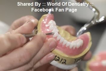
A reline is a procedure used to re-fit the surface of a denture to the tissues of the mouth.
This is done by placing a new acrylic base into the denture, duplicating the new form of the oral structures in the mouth.
A reline will bring back comfort to the patient, stability to the denture and discourage food from going underneath it.
Relines are required for many reasons with the most common reason being gum resorption (shrinking of the oral tissues).
Gum resorption is caused by the normal process of aging or after natural teeth have been extracted.
A general rule of thumb is that dentures should be relined once every 2 to 3 years.
Relines are a normal part of denture maintenance and important to the health of the oral tissues and bone support of a denture wearer.
A reline is not always recommended for a denture, If a denture has lost some retention and the "bite" or occlusion has changed considerably, a reline will not be satisfactory in correcting the retention of this denture.

-----------------------------------

Types of Relines:

A- Temporary Reline:
A temporary reline (tissue conditioner) is a silicone gel designed to last 1 to 4 months.
It is normally used in dentures of people who have had their natural teeth extracted or to improve the health of their tissues before new dentures are made.

B- Direct Reline:
A direct reline is also called a chair side reline.
The direct reline material is inserted in the denture and sets in the mouth as the patient waits in the dental chair.
The direct reline is offered in a hard or soft base, and are not meant as a long term solution to fit issues.

C- Processed Reline:
A processed reline is completed by taking an impression inside of the patient's existing denture duplicating the new form of the oral structures in the mouth.
This impression is then processed in our in-house laboratory for approximately 7 hours.
This is considered a permanent reline due to the high quality of material used and the precision of the fit achieved.

D- Soft Reline:
A soft liner is used mainly in full lower dentures and allows for more protection and comfort for people who have sensitive gums with little or no bone support.
The soft base is a liner that is bonded into your denture.
Your denture will adhere better to what remains of your gum, be less irritating to your tissues and give your denture more stability.
When constructing a denture, the Prosthodontist will take an impression for the soft base and process it into the inside of the denture.
The Soft liner material is a bonded medical grade rubberized base.
Soft liners will last approximately 2 years.
Remember: relines do not change the color or shape of the teeth or make them sharper for chewing, A reline replaces the fitting surface of the denture to make it fit better.

E- Rebase
A rebase is the process of refitting a denture by replacing the entire denture base.
This procedure is done by taking a new impression inside the denture, completely removing the old acrylic and adding a new acrylic base around the existing teeth.
This service is recommended for dentures that have had multiple repairs, discoloration or are very thin.

----------------------------

This Article has been Edited By :: World Of Dentistry :: TEAM
For any questions and suggestions please don't be hesitate to feedback us.

Yours,
:: World Of Dentistry :: TEAM

Thursday, August 15, 2013

Wilckodontics (Accelerated Osteogenic Orthodontics)


- Wilckodontics, also known as Accelerated Osteogenic Orthodontics, involves a periodontal procedure combined with orthodontics to reach end results of straight teeth 3 to 4 times faster than conventional orthodontic treatment.

- In the 1950s, periodontists were using corticotomy procedures to increase the rate of tooth movement. An oral corticotomy is a type of surgery where cuts are made in the jaw bone that surrounds and supports your teeth, called the alveolar bone.

- In the 1990s, the Drs. Wilcko, using CT scans, concluded that a marked reduction in mineralization of the Alveolar bone was the reason for the accelerated tooth movement following corticotomies.

- In 1995, Drs. Wilcko patented the AOO (Accelerated Osteogenic Orthodontics) technique.Unlike a usual corticotomy, AOO doesn’t just cut into the bone, but decorticates it.

- Brief about AOO steps:
1- That is, some of the bone’s external surface is removed.
2- The bone then goes through a phase known as osteopenia, where its mineral content is temporarily decreased.
3- The tissues of the Alveolar bone release rich deposits of calcium, and new bone begins to mineralize in about 20 to 55 days.
4- While the bone is in this transient state, braces can move your teeth very quickly, because the bone is softer and there is less resistance to the force of the braces.

-Research has shown that the results of AOO are as stable and long-lasting as conventional orthodontic braces. There seems to be more Alveolar bone present in patients who have undergone AOO, compared to before surgery, which is advantageous if your profile needs to be built up to improve your facial aesthetics.

----------------------------

Advantages of AOO:
1- Shortened treatment time (3-4 times).
2- Decreased chance of root resorption following orthodontics * Increased Alveolar bone providing better support for your teeth and facial profile.
3- Less likelihood for relapse.
4- You can choose from metal, ceramic, or gold brackets, or even Invisalign.
5- Since teeth are moving through a softened bone, there is less discomfort associated with teeth movement (following each orthodontic adjustment) compared to traditional orthodontics.

Disadvantages of AOO:
1- More expensive than conventional braces.
2- Being a mildly invasive surgery, it has its risks.
3- Expect some swelling and tenderness immediately after surgery.
4- Need to recover for a week and must stay away from school or work.

----------------------------

This Article has been Authored By :: World Of Dentistry :: TEAM
For any questions and suggestions please don't be hesitate to feedback us.

Yours,
:: World Of Dentistry :: TEAM

Monday, August 12, 2013

Branches of Maxillary nerve


Branches of Maxillary nerve

1. Middle meningeal nerve
2. Twiges to the sphenopalatine ganglion
3. Posterior superior alveolar nerve
4. Zygomatic nerve
5. Infra-Orbital Nerve
---- 5.A. Middle superior alveolar nerve
---- 5.B. Anterior superior alveolar nerve
---- 5.C. Terminal branches
--------- 5.C.1. Inferior palpebral nerve
--------- 5.C.2. External nasal nerve
--------- 5.C.3. Superior labial nerve

------------------------

3. Posterior Superior Alveolar Nerve
supplies
1- The Pulp
2- The Investing structures
3- The buccal mucoperiosteum of Upper Molars except MB root of 1st molar.

-------------------------

4. The Zygomatic Nerve
Divides into two branches:
1- Zygomatico-Tempopral
2- Zygomatico-Facial

--------------------------

5. The Infra-Orbital Nerve
Divides into:
1- The Middle Superior Alveolar Nerve
2- The Anterior Superior Alveolar Nerve
3- Terminal branches (Inferior Palpebral nerve - External Nasal Nerve - Superior Labial Nerve)

--------------------------

5.A. Middle Superior Alveolar Nerve
Supplies
1- Pulp
2- Investing structures
3- buccal mucoperiosteum of Premolars & MB root of 1st molar

--------------------------

5.B. Anterior superior alveolar nerve
Supplies
1- Pulp
2- Investing structures
3- labial mucoperiosteum of anterior teeth

--------------------------

- Sphenopalatine Ganglion
Braches into:
1- Orbital nerve
2- Nasal nerve
3- Palatine nerve
4- Pharyngeal nerve

--------------------------

- The greater (anterior) palatine nerve
Supplies palatal mucoperiosteum of maxillary molars & premolars

- The lesser ( posterior ) palatine nerve
supplies uvula, soft palate & posterior part of the hard palate

--------------------------

- Nasal Branches
1- Long sphenopalatine nerve (nasopalatine nerve) - supplies palatal mucoperiosteum of maxillary anterior teeth
2- Short sphenopalatine nerve

--------------------------

Read More About

Innervations of the Mandibular teeth
--------------------------

This Article has been Authored By :: World Of Dentistry :: TEAM
For any questions and suggestions please don't be hesitate to feedback us.

Yours,
:: World Of Dentistry :: TEAM

Innervations of the Maxillary teeth


Buccal Aspect: (pulp, investing structures and the buccal mucoperiosteum)

1- Posterior superior alveolar nerve supplies the maxillary third, second and the first molars except the mesiobuccal root of the first molar
2- Middle superior alveolar nerve supplies mesiobuccal root of the first molar, and the maxillary premolars
3- Anterior superior alveolar nerve supplies the anterior teeth

Palatal Aspect

1- The greater (anterior) palatine nerve It supplies the palatal mucoperiosteum opposite to the maxillary molars, premolars and canine.
2- The nasopalatine nerve It supplies the palatal mucoperiosteum opposite to the anterior region (including the canine)

Note: The accessory innervation of the Maxillary teeth is The upper anterior teeth cross innervation (Anterior Superior Alveolar Nerve crosses the midline from one side to another)

--------------------------

Read More About

Innervations of the Mandibular teeth

--------------------------

This Article has been Authored By :: World Of Dentistry :: TEAM
For any questions and suggestions please don't be hesitate to feedback us.

Yours,
:: World Of Dentistry :: TEAM

Innervations of the Mandibular teeth


Inferior Alveolar nerve
It supplies the pulp and the investing structures of the Mandibular molars, Premolars and Anterior teeth

Lingual nerve
It supplies the lingual mucoperiosteum of all the lower teeth, anterior two thirds of the tongue and the floor of the mouth.

The long Buccal nerve
It supplies the buccal mucoperiosteum opposite to the lower molars.

Note: The accessory innervation of the Mandibular teeth is
1- The lower anterior teeth cross innervation (Incisive Nerve crosses the midline from one side to another).
2- The lower premolar may receive additional nerve supply from the cutaneous coli nerve (branch from the cervical plexus of nerves) for their labial mucoperiostieum.
3- The lower molars may receive additional nerve supply from the nerve to mylohyoid for the pulp and investing structures.

--------------------------

Read More About

Branches of Mandibular nerve

--------------------------

This Article has been Authored By :: World Of Dentistry :: TEAM
For any questions and suggestions please don't be hesitate to feedback us.

Yours,
:: World Of Dentistry :: TEAM

Branches of Mandibular nerve


Innervations of the Mandibular Nerve

1. Main Trunk
2. Nervous Spimosum
3. Nerve to medial pterygoid
4. Anterior Division
---- 4.A. N. to Temporalis msc.
---- 4.B. N. to Massetter msc.
---- 4.C. N. to Lateral Pterygoid msc.
---- 4.D. Long Buccal N.
5. Posterior Division
---- 5.A. Auriculo-temporal N.
---- 5.B. Lingual Nerve
---- 5.C. Inferior Alveolar N.

---------------------------

Sensory braches of Mandibular nerve
1. Nervous spinosum nerve (trunk)
2. Long buccal nerve ( Ant. division)
3. Auriculo-Temporal nerve
---- 3.A- Parotid branch
---- 3.B- Articular branch
---- 3.C- Auricular branch
---- 3.D- Temporal branch
---- 3.E- Terminal branch
4. Lingual nerve ( post. division)
5. Inferior alveolar nerve (Incisive nerve - Mental nerve)
---- 5.A- Mylohyoid nerve
---- 5.B- Mental nerve
---- 5-C Incisive nerve

Motor braches of Mandibular nerve
1. Nerve to medial pterygoid muscle
2. Nerve to tensor palatini muscle
3. Nerve to tensor tympani muscle
4. Nerve to lateral pterygoid muscle
5. Nerve to masseter muscle
6. Nerve to temporalis muscle
7. Nerve to mylohyoid muscle
8. Nerve to anterior belly of digastric muscle

----------------------------

Long Buccal Nerve:
Supplies
1- M.M. of the cheek Except the posterosuperior area which receive sensory fibers from posterior superior alveolar nerve
2- Skin of the cheek
3- M.M. of the lower buccal vestibule
4- Buccal mucoperiosteum of the lower molars

The Lingual Nerve:
Supplies
1- M.M. of the ant. 2/3 of the tongue
2- M.M. of the floor of the mouth
3- M.M. of the lateral lingual vestibule
4- Lingual mucoperiosteum of the all lower teeth

Mylohyoid nerve:
Supplies:
1- Motor Fibers supplies the (Mylohyoid muscle - Anterior belly of digastric muscles)
2- Sensory fibers supply the (Skin of the anterior & inferior surfaces of the mental protuberance)

Mental nerve: supplies the (Skin of the chin - Skin of the lower lip - M.M. of the lower lip)

Incisive nerve: Supplies (pulp & investing structures of the lower anterior teeth)

--------------------------

Read More About

- Innervations of the Mandibular teeth
Branches of Maxillary nerve
- Innervations of the Maxillary teeth

---------------------------

This Article has been Authored By :: World Of Dentistry :: TEAM
For any questions and suggestions please don't be hesitate to feedback us.

Yours,
:: World Of Dentistry :: TEAM

Wisdom Teeth Pain and Other Common Symptoms


Why Do We Get Problems With Wisdom Teeth ?

Wisdom teeth pain symptoms, caused by their ‘impaction’, are a common problem. These ‘third molars’ are the last teeth to erupt into the mouth. There are usually four, one in each corner of the mouth at the back. They most commonly appear between the ages of 17 and 24, but can also erupt much later.
Symptoms are common here as they are the last molars to enter the mouth, there is often not enough space for them to fully come through. Therefore they may only partially erupt into the mouth or not come through at all.

When there is enough room, they will come through into the mouth normally and act as any other tooth. There may be some problems including dental pain as they are growing in, (you may thus occasionally need some mild pain relief – such as acetaminophen, paracetamol) but this will clear up once the tooth finds its final position.
Pain and other symptoms can arise when there is not sufficient room in the mouth. The tooth may become impacted ...

-------------------------------------

What is an Impacted Wisdom Tooth ?

A tooth is described as impacted if it is blocked from erupting into the mouth fully. 
Thus it will lie at an angle instead of being upright, remaining tipped against the tooth in front of it.
Technically, any tooth can become impacted but it is wisdom teeth that are the most often affected, owing to their late eruption.
Impacted teeth can cause a range of problems, but it should also be noted that they may cause no problems at all.

-------------------------------------

What Symptoms Might I Get ?

Minor Symptoms:

When a wisdom tooth is problematic symptoms may include:
- Pain and swelling of the gum overlying the impaction – this is due either infection of this operculum or trauma from the tooth above hitting into it, or a combination of both. For example, a swelling that arises from infection may make the upper tooth impinge onto the gum – traumatizing it more and causing a vicious cycle.
- Bad breath, due to infection and/or debris building up in the area.
- A bad taste in the mouth, for the same reasons.
- Pus coming out from the swollen gum area.
- Aches when you open your mouth, as you are stretching the inflamed tissues.
- Difficulty on opening your mouth.
- Tenderness when chewing or biting as this hurts the swollen gum area.
- Pain/ulcers on the inner cheek, where the pointy parts (cusps) of the impacted teeth may be digging into the soft tissues of the cheeks.
- Ear-ache, as pain can spread outward from the area.
The symptoms can occur for a few days and then clear up. It can then come back at any time, often with weeks or months between occurrences.

More Serious Symptoms:

More serious symptoms can develop:
Watch out for these signs, and note that they may develop quite quickly:
- Swollen glands under the chin (‘lymph nodes’).
- Swelling of the face and jaw, may indicate cellulitis.
- Muscle spasms in the jaw.
- Fever and general malaise.
- Such symptoms may indicate a severe, spreading infection which can be very serious if left untreated. - - --- Immediate advice should be sought from your dentist.

The cause of these problems is that when a wisdom tooth is impacted, a flap of gum will lie over it. As it is difficult to clean effectively under the gum flap, bacteria will proliferate here and the gum will become inflamed. This inflammation is known as ‘pericoronitis’.
Pericoronitis is usually relatively easy to remedy, as it usually remains localized. It is when it becomes a recurring problem (or if it ever gives rise to dangerous symptoms like those above) that extraction must be considered.

An impacted tooth can also be present in the mouth without you even knowing about it, because it may not be causing any symptoms. 
However other problems can also be associated with impacted wisdom teeth.
They are prone to decay. This is because food can trap around them and they are difficult to clean. 
The tooth lying beside will also be at increased risk of decay for the same reason.
The tooth may become sensitive and/or painful.
Likewise, the area is more prone to gum disease for similar reasons: it is difficult to clean.
Rarely, cysts and other such growths may form around an impacted tooth.
Therefore even when they aren't causing pain, or other noticeable symptoms, they should be checked regularly. 
Your dentist can make sure that all is well in the area, or if any damage begins to occur then the situation can be remedied sooner rather than later.

-----------------------------

Prevention of Symptoms:

The cleaner the area and your mouth in general is kept, the less likely that pericoronitis and other problems will occur.
Therefore general oral hygiene measures should be adhered to, including flossing in the area of the wisdom tooth and regular use of mouthwashes.
Your dentist can guide you on this.

----------------------------

Published By:
:: World Of Dentistry :: TEAM

Friday, August 2, 2013

Significant Lab Tests in Dentistry


Hematological Tests:
1- CBC: Complete Blood Count with platelets and WBC differential.
2- ESR: Erythrocyte Sedimentation RAte
3- P.T./I.N.R: Prothrombin Time/International Normalized Ratio
4- P.T.T: Partial Thromboplastin Time

Renal Assessment Tests:
1- Serum creatinine (S. Cr.)
2- Blood Urea Nitrogen (BUN)

Diabetes Assessment Tests:
1- FBS: Fasting Blood Sugar
2- PPBS: Post Prandial/ Post Meal Blood Sugar
1- HbA1C: Hemoglobin A1C

Liver Assessment Tests:
1- Hepatic Serology
2- Liver Function Tests (LFTs)

Bone Assessment Tests:
1- Serum Calcium (Ca2+)
2- Serum Phosphorus (PO4)
3- Alkaline Phosphate (AlkP)

HIV/AIDS Status Assessment Tests:
1- CD4 Count & Viral Load (HIV RNA)
2- CBC w/Pits. & WBC Diff.
3- LFTs: Liver Function Tests
4- PT/INR: Prothrombin Time/International Normalized Ratio

-------------------------------

This Article has been Authored By :: World Of Dentistry :: TEAM
For any questions and suggestions please don't be hesitate to feedback us.

Yours,
:: World Of Dentistry :: TEAM
Thursday, August 1, 2013

Schirmer's test


- Definition:
Schirmer's test determines whether the eye produces enough tears to keep it moist. 
This test is used when a person experiences very dry eyes or excessive watering of the eyes. 
It poses no risk to the subject. 
A negative (more than 10 mm of moisture on the filter paper in 5 minutes) test result is normal. 
Both eyes normally secrete the same amount of tears.
It is named for Otto Schirmer.

- Conditions:
Dry eyes can occur from conditions such as:
● Aging
● Dehydration
● Corneal ulcers and
● infections Eye infections (for example conjunctivitis)
● Vitamin A deficiency
● Sjögren's syndrome
● Secondary tearing deficiency (associated with disorders such as - lymphoma, leukemia, GVHD (graft vs. host disease, after a transplant), andrheumatoid arthritis)
● As a temporary or permanent side effect of LASER vision correction surgery such as LASIK or PRKThe inability of tears to drain into the nose can occur with:Some eye infections Blockage of the tear duct

-------------------------------

This Article has been Authored By :: World Of Dentistry :: TEAM
For any questions and suggestions please don't be hesitate to feedback us.

Yours,
:: World Of Dentistry :: TEAM

Monday, July 29, 2013

Management of excessive bleeding before or during surgery


Surgical Hemostatics = Styptics
1- Physical Methods: Pressure , Cold , Cautery.
2- Vasoconstrictors: Adrenaline.
3- Astringents: Tannic Acid & Alum (They Precipitate Blood Proteins Causing Contraction)
4- Local Coagulants. (To Be Discussed)
5- Systemic Coagulants. (To Be Discussed)

:: Local Coagulants ::
1- Thromboblastin. (Coagulin)
2- Thrombin.
3- Fibrin Glue.
4- Human Fibrin Foam.
5- Absorbable Gelatin Sponge.
6- Oxyce. (Oxidized Cellulose)

:: Systemic Coagulants ::
1- Fresh blood transfusion to restore blood volume and to supply coagulation factors.
2- IF DUE TO capillary fragility. Vit C + Vit P
3- IF DUE TO HEMOPHILIA. (Anti-Hemophilic Globulin Factor 8) or (Tranexamic Acid)
4- IF DUE TO FIBRINOLYRIC THERAPY. (Tranexamic Acid)
5- IF DUE TO ANTICOAGULANT THERAPY. (If heparin give protamine sulphate) , (If warfarian give vit K)

-------------------------------

This Article has been Authored By :: World Of Dentistry :: TEAM
For any questions and suggestions please don't be hesitate to feedback us.

Yours,
:: World Of Dentistry :: TEAM

Sunday, July 21, 2013

Dental Management of Patients taking Digitalis


Definition:
Digitalis is a Cardiac Glycoside drug prescribed for patients with Congestive Heart Failure (CHF) Or Atrial Fibrillation.

Mechanism of action of Digitalis:
- It binds and Inhibits the Magnesium and Adenosine Triphosphate Dependent Na+ and K+ ATPase.
- The increase the influx of calcium ions.
- Then it Enhances the myocardial contractility.

Contraindications:
- Local Anesthesia with Epinephrine.
- Aspirin (As it decreases Digitalis absorption and displaces it at the protien binding sites).
- NSAIDs (As they decrease renal clearance of Digitalis).
- Macrolides and Tetracycline (As they increase the serum levels of digitalis causing toxicity).

What is safe with Digitalis .... ?
- Anesthesia: Mepivacaine.
- Analgesics: Acetaminophen (Alone, with Codeine, with Hydrocodone Or with Oxycodone)
- Antibiotics: Penicillins, Cephalosporins and Clindamycin.

-----------------------------

This Article has been Authored By :: World Of Dentistry :: TEAM
For any questions and suggestions please don't be hesitate to feedback us.

Yours,
:: World Of Dentistry :: TEAM

Monday, July 15, 2013

Precision Attachment


- Definition:
It's part of a Dental Prosthetic Design that aims to connect a partial denture to fixed bridgework. can be used to restore arches where there are not enough teeth for fixed bridgework.

- Parts:
The Precision Attachment consists of two parts, Female and Male parts, the male part is attached to the partial denture, while the female part is soldered to fixed crowns or bridgework.

- Advantages:
1- Cosmetic Appearance.
2- Maintainable Periodontal Health.
3- Longevity of Abutment Teeth.
4- Patient Comfort.
5- Questionable Teeth can be saved in a way that does not affect case.
6- Longevity if they are lost in the future.
7- Natural Tooth and/or Implant Abutments can Be Used.
8- Can Be Adapted to Compensate for Future Changes in the Mouth.

- Function:
- There is no mechanical locking in the design of the Precision Attachment.
- A good precision attachment partial denture will not dislodge during normal function.
- The path of insertion of the male part inside the female part is different from the pull of the muscles and the action of the tongue and gravity, so that dislodging does not happen.
- It can move in a "vertical direction" slightly to release the forces instead of passing along them to the abutment teeth, The result is "physiologic stimulation" of the abutment teeth and the edentulous ridges.

- Requirements:
- All the teeth that support bridgework are prepared with full shoulder preparations in three-dimensions.
- Any defects must be corrected surgically, to create as healthy environment as possible.
- Minimize occlusal forces by: narrow occlusal (biting surface) diameters, adequate room for hygiene, proper length and anatomy, solid and passive fit, and adequate occlusion (bite) at the correct jaw relationship.
- Must be used in a precise manner in order to maintain a high percentage of longevity.
- Precision attachment cases must fit with precision.
- The abutments must be stable.
- The frameworks must fit properly without rock.
- Steps should be followed accurately without dismissing to avoid complications.

-----------------------------

This Article has been Authored By :: World Of Dentistry :: TEAM
For any questions and suggestions please don't be hesitate to feedback us.

Yours,
:: World Of Dentistry :: TEAM

Thursday, July 4, 2013

Clinical Periodontology and Implant Dentistry, 2008, 5th Edition, By Jan Lindhe


Description

The fifth edition of Clinical Periodontology and Implant Dentistry brings to its readers another iteration of the unrivalled, unparalleled work on the specialty of periodontics. The editors have brought together contributions from experts all over the world to provide the reader with a comprehensive, cohesive text that fuses scholarship and science with clinical instruction and pragmatism. With an increase in length of approximately 25% and 15 new chapters, the new edition of Clinical Periodontology and Implant Dentistry runs the gamut of sub-disciplines and topics within periodontics and implant dentistry, supporting an intellectually and internationally inclusive approach.

Review

“With so many excellent aspects of implant dentistry covered in such great detail, it should be easy to see why I therefore recommend this fine textbook as a ‘must reference’ for any practitioner involved with the placement and/or restoration of implants. The editors are to be commended for their studious and detailed fine work.” (Implant Dentistry, April 2009)

"I would highly recommend this book to undergraduates, postgraduates, clinicians and researchers … .Who will no doubt refer to this text time and time again." (Dental Update, November 2008)

"This is a comprehensive textbook … it is useful to those at all stages of their under-graduate career." (British Dental Journal, August 2008)

Clinical Periodontology and Implant Dentistry, 2003, 4th Edition, By Jan Lindhe


Description

This fourth edition of Clinical Periodontology and Implant Dentistry is the ultimate resource on periodontics. The editors are joined by over 50 expert contributors to address the full spectrum of periodontal issues. The book’s coverage extends logically from the anatomy of the periodontium through to treatment options, and elucidates the relationship between periodontal and restorative dental therapies. The section dedicated to implants in the periodontally-compromised patient reflects the increasing importance of this form of oral rehabilitation. This section includes new chapters on implant placement in the esthetic zone and issues related to supportive therapy in the implant patient.

With an approximate 25% increase in length, the fourth edition keeps pace with the level of interest and growth of research in periodontology. Chapters included in previous editions have been updated and many new chapters added, including modifying factors in periodontal disease, risk assessment, and genetics in relation to periodontitis.

The remarkable scholarship evident in the text is matched only by the quality of the illustrations, which guide the reader through the wealth of material detailed in the book. These features combine to make essential reading for the discerning student and practitioner and an asset to the faculty or practice library.

Key Features

Includes fourteen new chapters
Features 2000 illustrations, with over 1500 in color
Distills significant and original research findings
Includes detailed case reports
Editors and contributors provide an international perspective

Saturday, June 29, 2013

Russell-Silver Syndrome


- Definition:
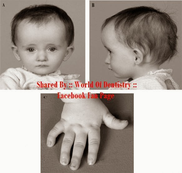
Russell-Silver syndrome is a disorder present at birth involving poor growth. One side of the body also will appear to be larger than the other.

- Causes:
Up to 10% of patients with this syndrome have a defect involving chromosome 7. In other patients, there the syndrome may affect chromosome 11, 15, 17, or 18.
Researchers suspect that at least one third of all cases of Russell-Silver syndrome result from changes in a process called methylation.
Methylation is a chemical reaction that attaches small molecules called methyl groups to certain segments of DNA.
In most patients, the cause is unknown. Most cases occur in people with no family history of the disease.

- Incidence:
The estimated number of people who develop this condition varies greatly. Some say it affects about 1 in 3,000 people. Other reports say it affects 1 in 100,000 people. Males and females are equally affected.

- Symptoms:
Body asymmetry
Coffee-with-milk (cafe-au-lait) birth marks
Curving of the pinky toward the ring finger
Delayed bone age
Failure to thrive
Gastroesophageal reflux disease
Kidney problems, such as:
Horseshoe kidney
Hydronephrosis
Posterior urethral valves
Renal tubular acidosis
Low birth weight
Large head for body size
Poor growth
Short arms
Short height (stature)
Short, stubby fingers and toes
Delayed stomach emptying, and constipation
Wide forehead with a small triangle-shaped face and small, narrow chin

- Signs:
Triangular shaped face in Russell-Silver syndrome patient
Pointed chin that is not fully developed
Thin, wide mouth
Triangle-shaped face with broad forehead

- Tests:
There are no specific laboratory tests to diagnose Russell-Silver syndrome.
Diagnosis is usually based on the judgment of your child’s pediatrician.
However, the following tests may be done:
- Blood Sugar (some children may have low blood glucose)
- Bone Age Testing (bone age is often younger than the child’s actual age)
- Chromosome Testing (may detect a chromosomal problem)
- Growth Hormone (some children may have a deficiency)
- Skeletal Survey (to rule out other conditions that may mimic Russell-Silver syndrome)

- Treatment:
- Growth hormone replacement may help if this hormone is lacking.
- Making sure the person gets enough calories, to prevent low blood sugar and promote growth
- Physical therapy, to improve muscle tone
- Special education, to address learning disabilities and attention deficit problems the child may have

- Prognosis:
Older children and adults do not show typical features as clearly as infants or younger children.
Intelligence may be normal, although the patient may have a learning disability.

- Complications:
- Chewing or speaking difficulty if jaw is very small
- Learning disabilities

------------------------------

This Article has been Authored By :: World Of Dentistry :: TEAM
For any questions and Suggestions please don't be hesitate to feedback us.

Yours,
:: World Of Dentistry :: TEAM

Petersons Principles of Oral and Maxillofacial Surgery - 2004 - 2nd Edition


Overview

Peterson's Principles of Oral and Maxillofacial Surgery 3rd Edition Download PDF Ebook. Michael Miloro encompasses a variety of diverse matters making it a novel text amongst the medical and dental specialties. It also presents color photographs of medical procedures, clear color diagrams and e-studying references. 

The aim of this concise, easy-to-learn two-quantity text is to supply an authoritative and at the moment referenced survey of the specialty of Oral and Maxillofacial Surgery. It contains the mandatory information for clinicians and is a perfect reference text for preparation for board certification within the specialty. It's also very effectively illustrated with over 2,000 shade pictures, figures, and drawings.

The anesthesia sections encompass comprehensive patient assessment and management in the peri-anesthetic period osseointegrated implant section to include chapters on comfortable and laborious tissue adjunctive procedures, zygomaticus and novum methods, in addition to a bit on implant prosthodontics for the surgeon. There are additionally new chapters on cleft orthodontics and distraction osteogenesis.

This book is of undoubted value for college students and recently certified practitioners of dentistry, for whom it acts as an entire and ready reference through which to achieve a firm foothold in oral and maxillofacial surgery.

Product Details

ISBN-13: 9781550092349
ISBN-10: 1550092340
Author: Michael Miloro
Edition: 2nd
Binding: Hardcover
Publisher: Pmph Usa
Published: June 2004

Saturday, June 15, 2013

Alveolar Osteitis "Dry Socket"


Definition:
- Inflammation of the alveolar bone (i.e. the alveolar process of the maxilla or mandible). Classically this occurs as a postoperative complication of tooth extraction.
- Occurs more commonly occur in the mandible than the maxilla, due to the relatively poor blood supply of the mandible and also because food debris tends to gather in lower sockets more readily than upper ones.
- It more commonly occurs in posterior sockets (molar teeth) than anterior sockets (premolars and incisors), where the created surgical defect is relatively larger, and because the blood supply is relatively poorer posteriorly.

Etiology:
This usually occurs where the blood clot fails to form or is lost from the socket, this leaves empty socket with bare bone with inflammation limited to the lamina dura "the bone which lines the socket".

Signs and symptoms:
- An empty socket, which is partially or totally devoid of blood clot.
- Bone may be visible or the clot may be filled with food debris which reveals the exposed bone once it is removed.
- Surrounding inflamed soft tissues may overgrow to cover the socket.
- Dull, aching, throbbing pain, which is moderate to severe.
- The pain may radiate to other parts of the head such as the ear, temple and neck.
- Pain starts from 2-4 days after extraction.
- Pain may persist for 10-40 days, but most commonly is 10-14 days with adequate treatment.
- The pain might be strong enough so that no medication can relief.
- Intraoral halitosis.
- Bad Taste.

Causes:
- Extraction site: Wisdom Teeth, specially the mandibular.
- Infection: Pre-existing infection in the mouth, such as necrotizing ulcerative gingivitis, chronic periodontitis or pericoronitis.
- Smoking: Smoking and tobacco use of any kind causes vasoconstriction of small blood vessels due to nicotine action.
- Surgical trauma: Excessive force or excessive tooth movements impair the repairing process causing the Alveolar osteitis.
- Vasoconstrictors: The use of Vasoconstrictors may increase the risk for alveolar osteitis, although it is used in local anesthetic solution to make a profound analgesia to the area, specially the areas of acute pain and inflammation.
- Oral contraceptives
- Radiotherapy: That decreases the blood supply to the area.

Diagnosis:
- Pain occurs 2-4 days after extraction.
- Usually part of a broken root or bone fragment is left in the socket.
- A dental radiograph (x-ray) may be indicated to demonstrate such a suspected fragment.

Prevention:
- Avoid excessive forces.
- Press on the alveolar plates to decrease the size of the socket opening.
- Instruct the patient to avoid cold food, beverages, toothpicks into socket, cleaning socket using the tongue or fingers and vigorous mouth rinsing.
- Rinsing with chlorhexidine (0.12% or 0.2%) or placing chlorhexidine gel (0.2%) in the sockets.
- Prophylactic antibiotics prior and after extraction for 3 days, although it's not recommended.
- Antifibrinolytic agents applied to the socket after the extraction.
- Debride the bony walls of the socket to encourage hemorrhage (bleeding) in the belief that this reduces the incidence of dry socket "there is no evidence to support this practice".
- Schedule extraction appointment for females taking oral contraceptives on days without estrogen supplementation (typically days 23-28 of the menstrual cycle).
- Scaling the teeth to be extracted prior to extraction.

Treatment:
1. Pain medication " Non-Steroidal Anti-Inflammatory Drugs".
2. Rinsing / cleaning the socket "removing any debris from the hole".
3. Applying medicated dressings. "antibacterials, topical anesthetics and obtundants, or combinations of all three"
4- Schedule a recall visiting every 3 days to rinsing and cleaning socket and changing the dressing.
5- Prescribe antibiotic to avoid infection of the socket.
6- Oral Hygiene Instructions.
7- Ask the patient to wash the mouth by water and salt or by prescribing Oral Mouthwash.

------------------------------

This Article has been Authored By :: World Of Dentistry :: TEAM
For any questions and Suggestions please don't be hesitate to feedback us.

Yours,
:: World Of Dentistry :: TEAM