Sunday, July 27, 2014

Blue Sclera "Definition and Diseases"


Definition:
Blue sclera is characterized by localized or generalized blue coloration of sclera because of thinness and loss of water content, which allow underlying dark choroid to be seen.

Diseases and Disorders:

1. Associated with high urine excretion:A. Folling syndrome (phenylketonuria)
B. Hypophosphatasia (phosphoethanolaminuria)
C. Lowe syndrome (oculocerebrorenal syndrome; chondroitin-4-sulfate-uria)

2. Associated with skeletal disorders:A. Brachmann-de Lange syndrome
B. Brittle cornea syndrome (blue sclera syndrome)-recessive
C. Crouzon disease (craniofacial dysostosis)
D. Hallermann-Streiff syndrome (dyscephalia mandibulooculofacial syndrome)
E. Marfan syndrome (dystrophia mesodermalis congenita)
F. Marshall-Smith syndrome
G. McCune-Albright syndrome (fibrosus dysplasia)
H. Mucopolysaccharidosis VI (Maroteaux-Lamy syndrome)
I. Osteogenesis imperfecta (van der Hoeve syndrome)
J. Paget syndrome (osteitis deformans)
K. Pierre Robin syndrome (micrognathia-glossoptosis syndrome)
L. Robert syndrome
M. Silver-Russell syndrome
N. Werner syndrome (progeria of adults)

3. Chromosome disorders:A. Trisomy syndrome
B. Turner syndrome

4. Ocular:A. Congenital glaucoma
B. Myopia
C. Repeated surgeries
D. Scleromalacia (perforans)
E. Staphyloma
F. Trauma

5. Miscellaneous:A. Ehlers-Danlos syndrome (fibrodysplasia elastica generalisata)
B. Goltz syndrome (focal dermal hypoplasia syndrome)
C. Incontinentia pigmenti (Bloch-Sulzberger syndrome)
D. Lax ligament syndrome
E. Minocycline-induced
F. Oculodermal melanocytosis (nevus of Ota)
G. Pseudoxanthoma elasticum (Grönblad-Strandberg syndrome)
H. Relapsing polychondritis

------------------------------------

This Article has been edited By :: World Of Dentistry :: TEAM
For any questions and suggestions please don't be hesitate to feedback us.

Yours,
:: World Of Dentistry :: TEAM

Friday, July 25, 2014

Indications and Contraindications of the Maryland Bridge


Indications:
• Periodontal Splinting
• Replacement of missing teeth
• Prosthodontic splinting
• Combination with Removable Partial Denture
• Changes in Occlusal table
• Strengthening Natural Teeth

Contraindications:
• Sensitivity to base metal alloys
• Changes in facial esthetic of abutments (Long spans - Inadequate enamel for bonding)
• Poor quality enamel
• Short clinical crowns
• Narrow embrasures
• Incisors with thin buccal lingual dimensions
• Pathologic occlusion (i.e.- bruxism)
• Malocclusion
• High caries rate or high caries risk

------------------------------------

This Article has been edited By :: World Of Dentistry :: TEAM
For any questions and suggestions please don't be hesitate to feedback us.

Yours,
:: World Of Dentistry :: TEAM

Saturday, July 19, 2014

Bronchial (Pharyngeal) Arches


:: 1st Pharyngeal Arch (also called "mandibular arch") ::


Muscular contributions
Muscles of mastication, anterior belly of thedigastric, mylohyoid, tensor tympani, tensor veli palatini

Skeletal contributions
Maxilla, mandible (only as a model for mandible not actual formation of mandible), the incus, and the malleus of the middle ear, also Meckel's cartilage

Nerve
Trigeminal nerve(V2 and V3)

Artery
Maxillary artery, external carotid artery

--------------------------------

:: 2nd Pharyngeal Arch (also called the "hyoid arch") ::
Muscular contributions
Muscles of facial expression, buccinator,platysma, stapedius, stylohyoid, posterior belly of the digastric 

Skeletal contributions
Stapes, temporal styloid process, hyoid (lesser horn and upper part of body), Reichert's cartilage 

Nerve
Facial nerve (VII)

Artery
Stapedial artery, hyoid artery

--------------------------------

:: 3rd Pharyngeal Arch ::
Muscular contributions
Stylopharyngeus

Skeletal contributions
Hyoid (greater horn and lower part of body), thymus, inferior parathyroids

Nerve
Glossopharyngeal nerve (IX)

Artery
Common carotid, internal carotid

--------------------------------

:: 4th Pharyngeal Arch ::

Muscular contributions
Cricothyroid muscle, all intrinsic muscles of soft palate (including levator veli palatini) except tensor veli palatini

Skeletal contributions
Thyroid cartilage, superior parathyroids, epiglottic cartilage
Nerve
Vagus nerve (X),superior laryngeal nerve

Artery
Right 4th aortic arch:subclavian artery
Left 4th aortic arch: aortic arch

--------------------------------
:: 6th Pharyngeal Arch ::

Muscular contributions
All intrinsic muscles of larynx except the cricothyroid muscle

Skeletal contributions
Cricoid cartilage, arytenoid cartilages, corniculate cartilage, cuneiform cartilages

Nerve
Vagus nerve (X), recurrent laryngeal nerve

Artery
Right 6th aortic arch: pulmonary artery
Left 6th aortic arch: pulmonary artery and ductus arteriosus


------------------------------------


This Article has been authored By :: World Of Dentistry :: TEAM
For any questions and suggestions please don't be hesitate to feedback us.

Yours,
:: World Of Dentistry :: TEAM

Thursday, July 3, 2014

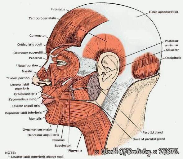
Muscles of the Face and Scalp

- Those muscles receive motor innervation from branches of the facial nerve (CN VII).

- Rather than inserting into bone, these muscles insert into the dermis of the skin, thus their orchestrated contractions convey various shapes to the face that we interpret as emotions.


Scalp

Muscle
Location
Origin
Frontalis
Forehead
Procerus, corrugator, orbicularis oculi
Occipitalis
Back of the head
Mastoid process and superior nuchal line
Temporoparietalis
Temple
Temporal fascia


Ear

Muscle
Location
Origin

Auricularis anterior
Anterior to ear
Temporal fascia
Auricularis superior
Above ear
Temporal fascia
Auricularis posterior
Behind ear
Mastoid process


Nose

Muscle
Location
Origin
Procerus
Nasalis
Depressor septi


Eye

Muscle
Location
Origin

Orbicularis oculi
Around the orbit
Nasal process of frontal bone, frontal process of maxilla, medial palpebral ligament, and lacrimal bone
Corrugator
Deep to the orbicularis oculi
Medial aspect of superciliary arch


Mouth

Muscle
Location
Origin
Levator labii superioris
Upper lip
Zygoma and maxilla just above infraorbital foramen
Levator labii superioris alaque nasi
Upper lip and side of nose
Maxilla, frontal process
Levator anguli oris
Corner of mouth
Canine fossa of maxilla
Zygomaticus major
Cheek and corner of mouth
Temporal process of zygoma
Zygomaticus minor
Cheek and corner of mouth
Maxillary process of zygoma
Risorius
Cheek
Masseteric fascia
Depressor labii inferioris
Lower lip
Oblique line of mandible
Depressor anguli oris
Corner of mouth
Oblique line of mandible
Mentalis
Chin
Incisive fossa of mandible
Orbicularis oris
Circumscribes the mouth
Muscles in the vicinity, maxilla, nasal septum, mandible
Buccinator
Cheek
Pterygomandibular raphe, alveola arches of mandible and maxilla


Neck

Muscle
Location
Origin
Platysma
Neck and chin
Pectoral and deltoid fascia


------------------------------------


This Article has been authored By :: World Of Dentistry :: TEAM
For any questions and suggestions please don't be hesitate to feedback us.

Yours,
:: World Of Dentistry :: TEAM